





 Canada
 UK
 France
 Australia
Most lymphedema referrals start the same way. A patient mentions a ring feels tighter, or a sleeve doesn't sit the way it used to, and by the time that gets reported, the fluid has usually been building for weeks. Tape measurement and clinical exam work well once swelling is visible, but neither of these are often strong enough to catch subclinical lymphedema. As a physical therapist and certified lymphedema therapist (CLT-LANA), I look for that earlier window, when fluid change is measurable but not yet something a patient notices or a tape measure detects.
Key Takeaways
- Subclinical lymphedema is a measurable increase in local tissue fluid that occurs before visible swelling or reported symptoms.
- Tape measurement and patient report are reliable for confirming established lymphedema, but neither is designed to detect a change that hasn't shown up yet.
- Risk factors and presentation differ by region: breast/chest, head and neck, and extremity lymphedema each have their own at-risk populations and their own reasons early change is easy to miss.
- Tissue dielectric constant (TDC) technology, the basis for LymphScanner, reads local tissue water content at a specific site and converts it to a percentage water content value, adding an objective data point to the standard exam.
Table of Contents
- What Is Subclinical Lymphedema and How Is It Detected?
- Why Tape Measurement and Patient Report Both Miss It Early
- Who's At Risk, and Why Presentation Differs by Region
- How TDC Technology Adds an Objective Data Point
- Where This Fits in the Plan of Care
What Is Subclinical Lymphedema and How Is It Detected?
Subclinical lymphedema is a measurable increase in local tissue water that occurs before lymphedema is visible or symptomatic. It's typically identified using objective, non-invasive measurement methods, such as tissue dielectric constant (TDC) technology, which reads local water content at a specific site rather than relying on visible swelling, patient-reported symptoms, or whole-limb measurements like circumference.
Why Tape Measurement and Patient Report Both Miss It Early
Circumferential measurement is a good tool for what it's built for: confirming lymphedema that's already established and tracking a known case over time. It's less useful for catching lymphedema at a subclinical level, before it reaches that visible threshold. Whole-limb volume measurement has the same limitation, because both methods track the size of the limb, and a small area of localized fluid may not change that size enough to detect.
The patient report has a similar limitation. Mild tightness or heaviness is easy to write off as normal post-surgical recovery, especially in the first several months, when some fluctuation is expected regardless. So although this is an important piece of a lymphedema therapist’s examination, it can’t be the only piece.
Who's At Risk, and Why Presentation Differs by Region
Breast and Chest
Patients who've had a mastectomy, lumpectomy, axillary node dissection, sentinel node biopsy, or chest wall radiation are all at increased risk for lymphedema. Early change here is easy to miss because normal post-surgical swelling and shifting garment fit look a lot like what you'd expect from healing.
Head and Neck
Neck dissection and radiation to the head and neck carry their own risk. Fluid change in this region tends to be subtle and diffuse, and there's no consistent landmark-based measurement comparable to what circumference offers for a limb.
Arms and Legs
Axillary or pelvic node dissection, radiation, and recurrent cellulitis all raise risk. Tape measurement works here, but as noted above, it tends to catch change only once it's already crossed a threshold.
How TDC Technology Adds an Objective Data Point
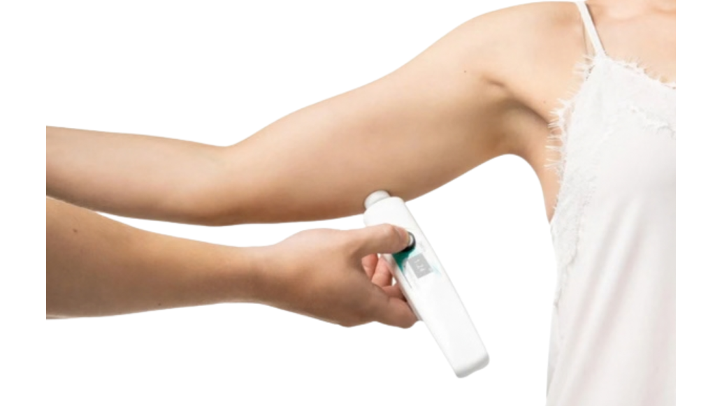
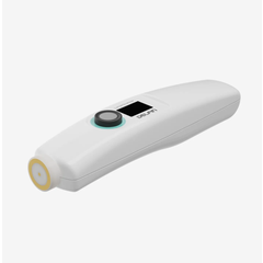
LymphScanner uses tissue dielectric constant (TDC) technology. A handheld probe placed against the skin sends a low-power electromagnetic wave into the tissue. Water has a high dielectric constant relative to fat and tissue proteins, so the reflected wave carries information about local water content.¹ The device converts that reading into a percentage water content (PWC) value, an objective number a clinician can track over time or compare between sites.
Because the reading is site-specific rather than averaged across a whole limb segment, it's designed to pick up localized changes that whole-limb methods, including circumference and volume measurement, aren't built to detect.² This matters most in the regions like the breast or head and neck, where there isn't a comparable manual alternative to begin with.
TDC vs. BIS: What Each One Measures
TDC and bioimpedance spectroscopy (BIS) are both used to assess lymphedema, and they measure different things. BIS sends a small current through the limb and estimates extracellular fluid across the whole segment between the electrodes. TDC reads water content in the skin and subcutis at a single site, roughly 2.5 mm deep. A limb can look normal to the first and abnormal to the second when the fluid is sitting superficially.
One study compared the two in 100 women within a year of breast cancer surgery, axillary dissection, and radiotherapy. Thirty-eight were clinically diagnosed with lymphedema. TDC identified more of them (sensitivity 65.8% vs. 42.1%), and 18.4% of the lymphedema cases were flagged by TDC alone, compared with 2.6% by BIS alone.² BIS was the more specific of the two (93.5% vs. 83.9%), so it produced fewer false positives. The authors attributed the difference to the tissue water each method assesses and concluded that nearly 20% of early lymphedema in this group was superficially localized.
That points to complementary tools. If BIS is already part of your workflow, TDC adds a look at the superficial compartment. If it isn't, TDC gives you a first objective measure.
Where This Fits in the Plan of Care
LymphScanner is FDA-cleared to support assessment of upper extremity lymphedema in adult women.
In your workflow, it's one more data point next to your history and exam findings. It doesn't diagnose on its own, and it doesn't replace your clinical judgment. Here's a practical way to build it in:
- Get a baseline. Measure before surgery or radiation when you can, or as early afterward as possible, so every later reading has the patient's own numbers to compare against.
- Use standardized sites. Pick the same landmarks on both sides at every visit and record them, so a change reflects the tissue and not where the probe landed.
- Compare sides and compare to baseline. Each site gives you a percentage water content value, so you can look at the ratio between the affected and unaffected side and at how a site trends over time.
- Read it alongside the rest of the exam. A rising number at one site tells you where to look harder. Check the skin, palpate, review symptoms, and take a tape measurement before you decide what it means.
- Re-test on a schedule. Keep visits closer together in the first two years after treatment, when risk is highest, and widen the interval after that.
Shop LymphScanner
References
- Mayrovitz, H. N. (2023). Medical applications of skin tissue dielectric constant measurements. Cureus, 15(12), e50531. https://doi.org/10.7759/cureus.50531
- Lahtinen, T., Seppälä, J., Viren, T., & Johansson, K. (2015). Experimental and analytical comparisons of tissue dielectric constant (TDC) and bioimpedance spectroscopy (BIS) in assessment of early arm lymphedema in breast cancer patients after axillary surgery and radiotherapy. Lymphatic Research and Biology, 13(3), 176–185. https://doi.org/10.1089/lrb.2015.0019
Medical Disclaimer: The information provided on this site, including text, graphics, images, and other material are for informational purposes only and are not intended to substitute for professional medical advice, diagnosis, or treatment. Always seek the advice of your physician or other healthcare professional with any questions or concerns you may have regarding your condition.